影像科中心

南华医院 影像科中心由顧問放射科醫生領導。還有四名顧問放射科醫師和一支合格的放射技師團隊為住院病人和門診病人提供診斷影像服務。 南華醫院配備* PACS,可無縫傳輸,檢索和存儲數字醫療影像,以改善病人的體驗。

*图片存档和通信系统 (PACS): 图片存档和通信系统(PACS)是一种用于存储,检索和传输数码医学图像的计算机网络系统。 PACS将来自系统的图像数据集成到系统中,允许在医疗保健设置内和之间进行传输。这有助于在需要时在护理点处提供图像和图像相关数据。


目标

为病人护理提供有效,高效和全面的诊断服务。


营业时间:周一至周五上午8时30分至下午6时
 周六上午8时30分至下午2时
 周日与公共假期休息
 
联络方式:电话号码:
 
地点:最底层

我们最先进的数码放射成像系统可提高生产率,病人舒适度和工作流程。生成的图像可以通过网络在整个医院进行数字化传输,以改善病人的周转时间和吞吐量。

  • Chest X-Ray
  • Skull X-Ray

荧光透视是提供实时X射线成像的方法,其对于指导各种诊断和程序特别有用。透视显示运态的能力是由连续的一系列图像造成。这类似于传统电视或视频传输图像的方式。在荧光检查过程中,X射线束穿过身体。将图像传输到监视器,以便可以详细地看到身体部位或造影剂(“染料”)穿过身体的运动。

荧光透视如何进行?

  • 通常整个检查需要30分钟到1小时
  • 消化系统
    钡吞咽/钡餐:涉及吞嚥钡以勾画胃肠道。
  • 泛影葡胺灌腸:泛影葡胺通過肛門途徑引入大腸。
  • 泌尿系统
    将小导管引入泌尿道并引入造影剂以显现膀胱和下泌尿道。
  • 生殖系统
    通常使用通过阴道和子宫颈注入子宫腔的造影剂进行测试。
  • 放射科医师将上下移动探测器,以便最好地查看正在检查的区域,图像将显示在监视器上。

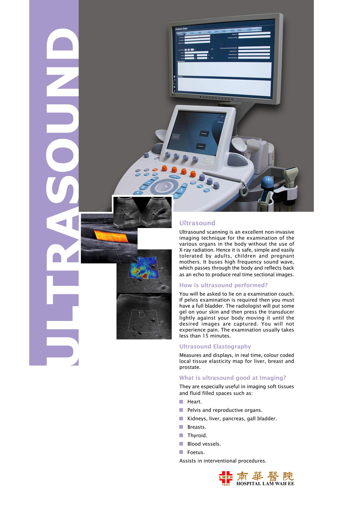
超聲掃描是一種優秀的非侵入性成像技術,用於在不使用X射線輻射的情況下檢查身體中的各種器官。因此它屬於一種安全的技術,成人,兒童和孕婦容易忍受。它匯集高頻聲波,通過身體反射回來作為迴聲,產生實時截面圖像。

超声波怎样进行?
你将被要求躺在检查沙发上。如果需要骨盆检查,那之前您必须忍尿直到您的膀胱被填满。放射科医生会在您的皮肤上涂上一些凝胶,然后轻轻按压传感器,将其移动到您的身体上,直到拍下所需的图像。你不会经历疼痛。检查通常不到15分钟。

超声弹性成像
实时测量和显示肝脏,乳房和前列腺的彩色编码局部组织弹性图。

为什么超声波对成像由益?
  • 它有助于诊断涉及肾脏,肝脏,胆囊,脾脏和胰腺的腹部疾病。
  • 它提供了表面组织的详细信息,如乳房,甲状腺和一些肌肉骨骼系统
  • 它允许准确评估盆腔区域充满液体的空间
  • 测量血管中的血流以检测异常

电脑断层扫描是一种无痛程序,它结合了数码电脑和旋转X射线器的使用,以创建详细的横截面图像或器官和身体部位的“切片”。

为什么要进行电脑断层扫描?
电脑断层扫描可以为身体的几乎每个部位提供详细的横断面图像和诊断资料,包括大脑,颈部,胸部,嵴柱,腹部,骨盆,心脏和骨骼系统。

准备及其执行方式
电脑断层扫描是一种非常安全的检查,是一种门诊检查。根据检查的类型,手术的时间通常需要10到45分钟。实际曝光/扫描时间非常短(5到10秒)。

对于许多人来说,电脑断层检查可能需要特殊的造影剂(口服,直肠或注射),有时也称为“染料”。 电脑断层对比用于製作特定器官;血管和/或组织类型“突出”具有更多图像对比度以更好地显示疾病或损伤的存在。

什么是磁力共振成像?
磁力共振成像是一种使用强磁场创建身体截面图像而无需使用X射线或辐射的方法。 磁力共振成像机器通过组织和操纵磁场内的水分子来製作图像。 磁力共振成像图像显示身体的软组织(肌肉,神经,脑盘,韧带等)比常规X射线和电脑断层扫描更清晰。

在大多数情况下,您可以在扫描进行前后进食和饮水。你会被问到: -
  • 在进入扫描室之前必须移除任何金属物体(手錶,钥匙,硬币,信用卡,髮夹等)
  • 在手术过程中必须躺着不动。

您可能会在手术过程中听到砰砰声。

如果您有任何以下物品,进行磁力共振扫描可能不安全(请谘询您的医生):
  • 心脏起搏器
  • 大脑中的手术夹(动脉瘤夹)
  • 人工心脏瓣膜
  • 电子模拟器
  • 内耳植入物

如何减少对图像造成的影响?
请告知放射技师如果您有
  • 牙科填充物和牙桥
  • 髋关节和膝关节置换术
  • 输卵管结扎手术
介入性放射学是放射学的一个分科,其中使用图像引导进行微创手术。这些程序中的一些是为了纯粹的诊断目的(例如,血管造影)而进行的,而其他程序是为了治疗目的而进行的(例如,血管成形术)。图片(图像)用于指导这些程序,这些程序通常使用针或其他微小仪器(如称为导管的小管)来完成。这些图像提供了路线图,允许介入性放射科医生将这些器械引导穿过身体到涉及的区域。
常见的介入性成像模式包括透视,计算机断层扫描(电脑断层),超声(US)和磁力共振成像(MRI) )。

牙科成像(OPG-正视图,侧位头影测量和CBCT-锥形束计算机断层扫描)

多年来,牙科博爱一直依赖于二维成像(Orthopantomography和Lateral Cephalometric),现在是我们转向三维CBCT技术的时候了。

我们的设备能够生产二维牙科造影和3-D CBCT体积数据。 2-D或3-D的使用在很大程度上取决于要回答的临床问题。

3-D CBCT图像可以帮助我们的牙科医生更好地诊断和了解口腔疾病的有效性并在口腔区域提供更合适的治疗。通过在一次扫描中区分解剖结构和多种类型的组织,体积数据的多平面视图提供了巨大的优势。

Mammogram (Digital Breast Tomosynthesis)

Mammogram is a special x-ray examination of the breasts. It is a recommended screening test for breast cancer. It can detect cancer at its early stage before it can be felt by hands. Early detection means better survival rate and more treatment options.

We recently installed a new mammography machine with digital breast tomosynthesis (DBT) capability which by using the cutting edge technologies to provide a more detailed sectional (3D) assessment of the breasts.


Mammogram procedure

  • You will be positioned and your breast will be placed on a special platform.
  • The breast will be compressed with a clear plastic compressor.
  • X-ray is exposed to produce an image of breast tissue.
  • To capture different views of breast, position is changed slightly between images.
  • The process is repeated for the other breast.
  • Some discomfort may be felt when the compressor compresses your breast. You can postpone the procedure until your breast is less tender (generally after your menses).

Before a mammogram

  • Do not wear deodorant, talcum powder or lotions under your armpit or around your breasts.
  • Inform radiographer if you have any symptoms or history.
  • Bring along previous mammogram films for comparison.
  • You are required to remove all your jewellery

You are advised to have your mammogram done if you have any of the following:

  • 40 years old and above.
  • Last mammogram was 2 years ago.
  • Family history of breast cancer.
  • Menopause and taking harmone replacement therapy.
  • You have history of breast cancer.
  • You feel lumps or there are some abnormal changes in your breasts recently.
  • Feel free to talk to your doctor about mammogram or call 04-652 8985 for further assistance.

Click here for details.

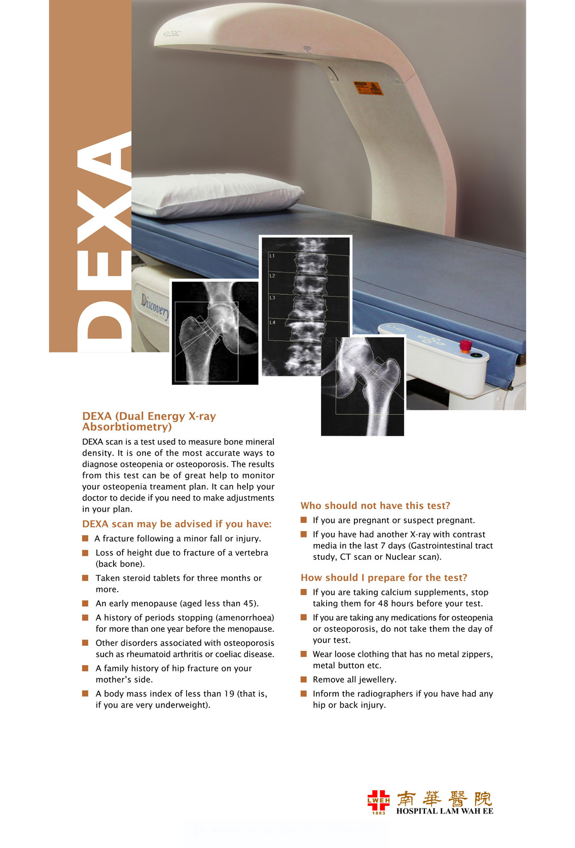
DEXA (Dual Energy X-ray Absorbtiometry)

DEXA Scan is a test used to measure bone mineral density. It is one of the most accurate ways to diagnose osteopenia or osteoporosis. The results from this test can be of great help to monitor your osteopenia treatment plan. It can help your doctor to decide if you need to make adjustments in your plan.

DEXA Scan may be advised if you have:

  • A fracture following a minor fall or injury
  • Loss of height due to fracture of a vertebra (back bone)
  • Taken steroid tablets for three months or more
  • An early menopause (aged less than 45)
  • A history of periods stopping (amenorrhoea) for more than one year before the menopause
  • Other disorders associated with osteoporosis such as rheumatoid arthritis or coeliac disease
  • A family history of hip fracture on your mother's side
  • A body mass index of less than 19 (that is, if you are very underweight)

Who should not have this test?

  • If you are pregnant or suspect pregnant
  • If you have had another X-ray with contrast media in the last 7 days (Gastrointestinal tract study, CT scan or nuclear scan)

How should I prepare for the test?

  • If you are taking calcium supplements, stop taking them for 48 hours before your test
  • If you are taking any medications for osteopenia or osteoporosis, do not take them the day of your test
  • Wear loose clothing that has no metal zippers, metal button etc.
  • Remove all jewellery
  • Inform the radiographers if you have had any hip or back injury

Radiologists
放射学科医生

Dr Ho Chee Keong   何智强医生

MMC : 27610 / NSR: 124019

Imaging Centre, Ground Floor

Dr Lee Cheng Hock   李清福医生

MMC : 22872 / NSR: 124020

Imaging Centre, Ground Floor

Dr Chan Kam Hong   陈锦丰医生

MMC : 43296 / NSR: 131311

Imaging Centre, Ground Floor

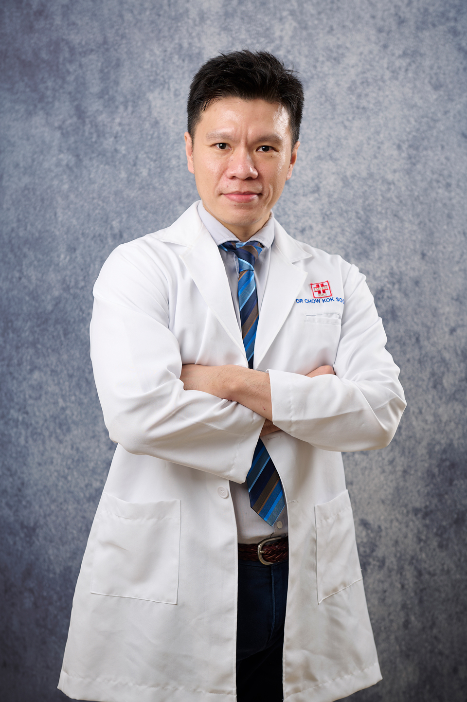
Dr Chow Kok Soon   周国顺医生

MMC : 45850 / NSR: 132793

Imaging Centre, Ground Floor

Dr Tan Yuan Ying   陈瑗溵医生

MMC : 47947 / NSR: 138648

Imaging Centre, Ground Floor

Dr Ang Lee Nah   洪丽娜医生

MMC : 49438 / NSR: 139537

Imaging Centre, Ground Floor


联络方式

位置
No. 141, Jalan Tan Sri Teh Ewe Lim, Jelutong, 11600, Pulau Pinang, Malaysia
+604 - 652 8888